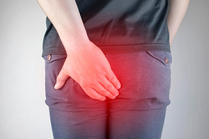
Perianal masses
Perianal masses
This article was updated in January 2026.
Perianal masses are a common presentation in primary care, and can cause significant anxiety (not to mention discomfort) for our patients. The causes can vary from minor (haemorrhoids) to much more serious (perianal cancers). Treatment for perianal infections is often surgical so it is useful to be aware of the common presenting features and baseline management for the most common causes we might see.
In this article, we review the common causes of perianal masses and then consider some of these causes in more detail. We have drawn on the 2020 European Society of Coloproctology guidance on the management of haemorrhoidal disease, alongside articles from the BMJ, BJGP and American Family Physician, plus a healthy dose of Red Whale common sense!
|
Unexplained anal mass: consider referral on a suspected cancer pathway (NICE 2015 (updated 2023) NG12). Patients with an anal or rectal mass, or ulceration, do not require FIT prior to referral. |
Common causes of a perianal mass
| Cause of lump | Presenting features |
| Perianal abscess |
Usually presents as a tender, localised swelling with redness. Deeper abscess can present with vague perianal or pelvic pain (BMJ 2017;356:j475). Some may have discharged by presentation. Almost all require surgical drainage. Antibiotics have not been shown to improve outcomes. |
| Pilonidal sinus |
A lump in the natal cleft which is usually red, hot and tender, and may exhibit chronic drainage of pus (BJGP 2024;74:44). Management of pilonidal sinus is surgical: urgent if acute and routine if recurrent but currently not symptomatic. Do not delay referral to trial oral antibiotic treatment. |
| Haemorrhoids |
Bleeding is the most common symptom of haemorrhoids, and haemorrhoids are the most common benign cause of anal bleeding (Am Fam Physician 2020;101:24). The mass from a haemorrhoid may present as (Aus Fam Physician 2010;39:376):
|
| Anal cancer |
An ulcerated or indurated lesion with possible symptoms of pain, bleeding, changes in bowel habit and weight loss (Am Fam Physician 2018;97:172). Anal cancer is most commonly squamous cell carcinoma, and 90% are attributable to HPV (Surg Oncol Clin N Am 2017;26:57). Refer via suspected cancer pathways. |
| Skin conditions |
Common anal skin conditions that may present with lumps include (Am Fam Physician 2020;101:24):
We have separate articles covering genital warts and HPV, molluscum contagiosum and hidradenitis suppurativa. |
| Rectal prolapse |
Mass protruding through the anus after straining (Am Fam Physician 2020;101:24).
|
Perianal abscess
This is summarised from a BMJ review (BMJ 2017;356:j475).
Epidemiology
- Incidence around 16/100 000. Result in around 12 500 NHS operations a year.
- Mean age 40y.
- Twice as common in men as women.
- Most are idiopathic. Known risk factors include: inflammatory bowel disease, smoking, HIV. Rarely caused by abdominal or pelvic infection (e.g. diverticulitis), anal trauma or anal cancers.
- NOT related to personal hygiene or sedentary lifestyle.
Clinical features
- Superficial abscesses usually present as tender, localised swelling with redness. Some may have discharged by presentation.
- Deeper abscesses can be hard to diagnose – they take longer to present and may cause vague pelvic or perianal pain. Imaging may be needed.
- Rarely, perianal abscesses present with sepsis.
- Fistulas:
- Fistulas are usually the result of an abscess and only rarely occur alone.
- In one-third of people with an abscess, a fistula is present either at the time of presentation or after drainage. The exception to this is when there is an underlying cause (inflammatory bowel disease, TB, trauma, post surgery).
- There is no way of knowing who will develop a fistula, or of preventing its occurrence.
Management
-
Surgery: almost all perianal abscesses require drainage, even if they have spontaneously discharged. Surgery is usually done within 24h.
- Incision and draining can be under general or local anaesthesia. Most are drained externally, but very deep abscesses may be drained into the anal canal.
- The cavity should heal by secondary intention, so wounds may be packed or left open (in which case, the patient may be taught ‘digitation’ – rubbing the base of the wound with their finger). Packing increases pain scores and costs, and in a small RCT was not associated with better outcomes.
- About 8% will need re-operation within 7d, usually because of incomplete initial drainage, premature skin closure or missed loculations.
- Antibiotics: use of antibiotics before or after drainage does not seem to improve healing rates or reduce recurrence in uncomplicated disease. Antibiotics may be used in those with systemic symptoms, extensive cellulitis or underlying immunosuppression.
- General examination and sigmoidoscopy may be indicated if there is evidence of co-existing disease (inflammatory bowel disease, malignancy). Faecal calprotectin may be measured if there is concern about inflammatory bowel disease.
-
Post-operatively, the wound should be monitored for:
- Worsening symptoms.
- Persistent or spreading cellulitis.
- Malaise or pyrexia.
- If people are not making progress as you would anticipate, serial inflammatory markers may be useful, although clearly re-referral may be indicated in this situation (there could be a deeper abscess, or the skin may have closed over too soon after surgery, allowing pus to recollect (personal communication with author)).
Follow-up
- Follow-up is NOT needed (once it heals) if this is a first perianal abscess in the absence of systemic disease/underlying cause.
- Those with recurrent disease do need investigation and follow-up.
Pilonidal sinus
A pilonidal sinus is not a perianal abscess. However, the conditions are related, with similar risk factors and comorbidities. In perianal abscess, the natal cleft will be clear.
The management of pilonidal sinus was reviewed in a BJGP Clinical Practice article (BJGP 2024;74(738):44).
Ingrowth of a hair in the natal cleft leads to formation of a sinus, with a hair at the apex. The sinus is unable to drain, leading to abscess formation, with pain and chronic drainage of pus. Patients may have difficulty walking or sitting, and there may be excessive granulation tissue at the site.
Incidence is 26 per 100 000 and is more common in males than females (3:1 ratio).
Management is with drainage by emergency surgery. We should not delay referral for surgical care to try oral antibiotics. In those with chronic symptoms, referral to colorectal services for more extensive flap repair may be necessary.
Haemorrhoids
Epidemiology
Haemorrhoidal disease is common. In one study of patients attending for routine colorectal cancer screening, prevalence was found to be 38–39%, although exact numbers are hard to quantify because many are asymptomatic or do not seek medical advice (Am Fam Physician 2018;97:172, JAMA 2025;334:1545).
Haemorrhoids are more common between the age of 45 and 65 years (Am Fam Physician 2018;97:172). Association is seen with an increased pressure in the haemorrhoidal venous plexus, for example due to constipation, pregnancy, obesity, pelvic floor dysfunction, anal intercourse or chronic diarrhoea.
Clinical features
Typical presenting symptoms include (Am Fam Physician 2018;97:172):
- Bright red rectal bleeding: streaks of blood on the stool.
- Itch.
- Grape-like lumps protruding from anus.
- Pain may occur with external or thrombosed haemorrhoids.
Examination
The European Society of Coloproctology recommends examination of the anorectal region with the patient in the left lateral position (if comfortable for them) to exclude other localised pathology (Colorectal Diseases 2020;22:650).
External haemorrhoids may be visible in the left lateral, right anterior or right posterior aspect of the anus (Am Fam Physician 2018;97:172).
A rigid anoscope, proctoscope or rectoscope should be used to visualise the entire anal canal to exclude other pathology and classify the severity of haemorrhoids (Colorectal Diseases 2020;22:650).
Management
Initial management should include (Colorectal Diseases 2020;22:650):
- Dietary advice: high-fibre diet, sufficient water intake.
- Physical activity to promote bowel transit.
- Toileting advice: avoid straining, adopt ‘correct body position’ (the Society of Coloproctology doesn’t explain what it means by this, but we suspect it means hip flexion in a squatted position!).
- Laxatives: can be used for symptom relief and to reduce bleeding, despite a low level of evidence of benefit.
- NSAIDs and non-opioid analgesics can be prescribed for pain (although the BNF recommends avoiding NSAIDs if bleeding is present).
- Phlebotonics: a class of drug found to be beneficial for symptoms of bleeding and pruritus in a 2012 Cochrane review (Cochrane Database Syst Rev 2012, CD004322). These drugs are made up of plant extracts such as flavonoids. Their precise mode of action is not known, but they are associated with improved venous tone and lymphatic drainage.
Topical treatments with corticosteroids and local anaesthetics may help with local inflammation, but there is no evidence that they reduce swelling, bleeding or protrusion of the haemorrhoids. Use beyond 7 days should be avoided due to risk of skin damage (BNF, accessed January 2025).
Surgical treatment
If initial management fails or symptoms are recurrent, we should consider referral for surgical management. This may include rubber band ligation, injection sclerotherapy, photocoagulation or haemorrhoidectomy (JAMA 2025;334:1545).
The European Society guidance suggests that rubber band ligation would be the preferred choice for more minor haemorrhoids, with haemorrhoidectomy reserved for more severe cases, but that the choice of procedure should be based on shared decision-making, taking into account patient preferences, availability of procedures and patient frailty.
Thrombosed haemorrhoids
Despite a very limited evidence base, the consensus in the European Society of Coloproctology guidelines was that surgical treatment should be considered on a case-by-case basis.
A review found that excision of a thrombosed haemorrhoid within 72h of first onset results in (Am Fam Physician 2020;101:24):
- Faster symptom resolution (3.9 days compared with 24 days for conservative management).
- Reduced recurrence rates: 6% (surgery) vs. 25% (conservative) at one year.
|
Perianal masses
|
This information is for use by clinicians for individual educational purposes, and should be used only within the context of the scope of your personal practice. It should not be shared or used for commercial purposes. If you wish to use our content for group or commercial purposes, you must contact us at sales@red-whale.co.uk to discuss licensing, otherwise you may be infringing our intellectual property rights.
Although we make reasonable efforts to update and check the information in our content is accurate at the date of publication or presentation, we make no representations, warranties or guarantees, whether express or implied, that the information in our products is accurate, complete or up to date.
This content is, of necessity, of a brief and general nature, and this should not replace your own good clinical judgment or be regarded as a substitute for taking professional advice in appropriate circumstances. In particular, check drug doses, side effects and interactions with the British National Formulary. Save insofar as any such liability cannot be excluded at law, we do not accept any liability for loss of any type caused by reliance on the information in these pages.
Here is the link to our terms of use.